Agricultural sciences
June 28, 2023
Development of Technologies for Automatic Measurement and non-destructive observation of Stomata in Arabidopsis thaliana
【Key Points of This Study】
・Development of an image analysis algorithm for automatic quantification of stomatal aperture in Arabidopsis thaliana
・Development of a portable imaging device for non-destructive observation of stomata using intact plants
・These technologies allow for rapid and reliable measurement of subtle changes in stomatal aperture in response to light, chemicals, and microorganisms
【Research Overview】
A team of researchers led by Drs. Momoko Takagi and Yosuke Toda (concurrently affiliated with phytometrics co. ltd.) at the Institute of Transformative Bio-Molecules (WPI-ITbM※) at Nagoya University, part of the Tokai National Higher Education and Research System, in collaboration with Drs. Rikako Hirata and Akira Mine at the Graduate School of Agriculture, Kyoto University, have developed an image analysis algorithm for automatic quantification of stomatal aperture in Arabidopsis thaliana, as well as a portable imaging device for non-destructive observation of stomata using intact plants.
Stomata are pores on the surfaces of plant leaves that open or close in response to changes in the environment. AlthoughArabidopsis thaliana is a widely used model organism for studying the role of stomata in plant environmental responses, measuring stomatal aperture in this model plant with small stomata is a daunting task.
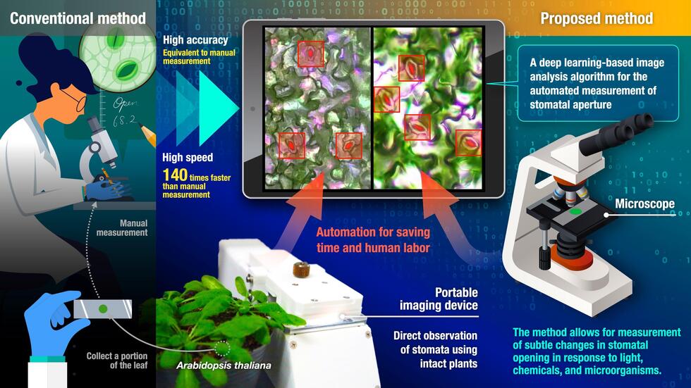
The research team has developed an image analysis algorithm that can automatically measure A. thaliana stomatal aperture, taking 140 times less time than manual measurement while maintaining the same accuracy. The research team also developed a portable imaging device that can capture stomatal images by pinching a leaf with the help of image acquisition software, making it possible to observe stomata using intact plants. These technologies allow for rapid and reliable measurement of subtle changes in stomatal aperture in response to light, chemicals, and microorganisms. The research results were published online in the journal "Plant & Cell Physiology" on March 20, 2023.
【Research Background and Result】
Stomata are pores surrounded by a pair of guard cells on the surface of plant leaves. Plants regulate the uptake of carbon dioxide needed for photosynthesis and the transpiration of water by controlling the degree of stomatal opening, also known as stomatal aperture. Plants prevent microbial invasion by closing stomata when detecting microbial molecules. Therefore, the measurement of stomatal aperture is essential to understand plant environmental adaptation. However, acquiring stomatal images under a microscope and dealing with a large number of stomata is time-consuming and labor-intensive. This is especially challenging for A. thaliana, a widely used model organism for studying the role of stomata in plant environmental responses, due to its extremely small stomata.
Traditionally, leaf discs (Note 1) have been widely used for microscopic observation of stomata, but the process of making leaf discs can affect stomatal aperture. Therefore, it is necessary to directly observe stomata on the leaf surface of intact plants to accurately evaluate stomatal responses. Many researchers have called for methods to enable such observations.
This study reports the establishment of an image analysis method that uses machine learning to automatically quantify A. thaliana stomatal aperture, as well as the development of a portable imaging device that can easily capture stomatal images by pinching a leaf of the intact plant at the site where the plant is growing.
Construction of Datasets and Machine Learning Models
Leaf discs are commonly used for experiments that measure stomatal aperture, but these microscopic images are difficult to analyze due to “noise” such as epidermal cells and mesophyll cells, making it difficult even for the human eye to locate stomata. Moreover, stomatal aperture changes dynamically in response to various environmental stimuli. Therefore, the research team collected microscopic images of A. thaliana stomata showing various apertures by exposing leaf discs to different light conditions and chemical compounds, and, annotated the information about the position, opening, and opening area of the stomata, with the help of researchers experienced in stomatal aperture measurement. These images were divided into training, validation, and test datasets, and used for developing machine learning models that can automatically measure A. thaliana stomatal aperture.
The research team developed a two-stage deep learning algorithm composed of object detection and region segmentation. Object detection retrieves the coordinate information of stomata and classifies them as open or closed. Various models of You Only Look Once X (YOLOX) (Note 2) were trained using the aforementioned training dataset, and the model YOLOX-s, which showed high accuracy and processing speed for the validation dataset, was selected. In the subsequent region segmentation, the opening area of the stomata determined to be "open" by YOLOX-s is detected and measured. The research team selected a model based on U-Net that showed the highest performance in detecting stomata and extracting the opening area for the validation dataset. The developed deep learning algorithm enabled automatic measurement of stomatal aperture in leaf disc images with an error of only 0.2 µm ± 0.2 µm compared to manual measurement (Figure 1). Furthermore, a detailed comparative analysis using the test dataset proved that the deep learning algorithm developed can capture changes in stomatal aperture in response to light and chemical compounds with equivalent accuracy to manual measurements.
Figure 1: Representative results of automatic stomatal aperture measurement (left),
and a researcher using the developed algorithm (right)
Portable Stomatal Imaging Device
Leaf discs can be easily prepared and therefore are practical samples for observing stomata using a microscope. However, it has been found that the damage caused by cutting the leaf from the plant and the accompanying production of plant hormones can affect stomatal aperture. Therefore, to precisely evaluate the physiological responses of stomata, it is necessary to directly observe stomata on a leaf of an intact plant.
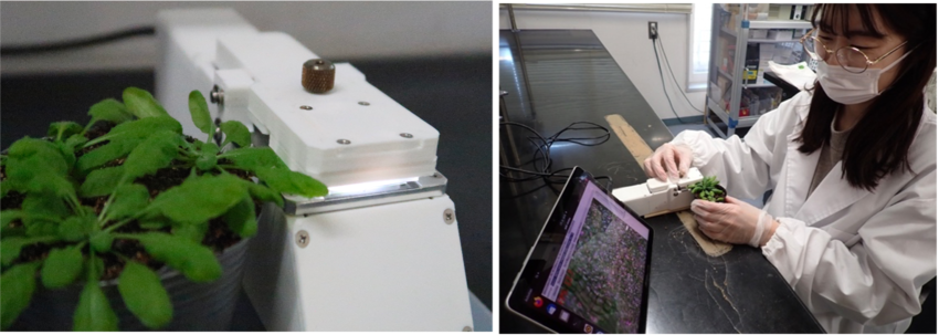
The research team developed a device that can capture images of stomata by pinching leaves of intact A. thaliana plants. This device is 5 cm wide × 20 cm deep × 6 cm high, and weighs 310 g, making it highly portable (Figure 2). However, the researchers noticed that the deep learning algorithm trained with microscopic images did not show reasonable performance with images captured by the portable stomatal imaging device. To address this issue, the research team retrained the model using images captured by the portable stomatal imaging device, obtaining a fine-tuned model that is able to detect stomata and measure their aperture with high accuracy for images captured by the portable stomatal imaging device. Furthermore, by combining the portable stomatal imaging device with the deep learning algorithm, the researchers successfully analyzed the stomatal response of A. thaliana to the bacterial pathogen Pseudomonas syringae. Notably, the time required for stomatal aperture measurement was reduced from an average of 211 seconds per image for manual measurement to 1.5 seconds for automatic measurement, achieving a 140-fold reduction in processing time.

Figure 2: Portable stomatal imaging device (left)
and an example of measuring stomatal aperture using the device (right)
Significance
This study introduces an image analysis algorithm that automates the measurement of A. thaliana stomatal aperture, achieving the same accuracy as manual measurement but in 140 times less time. Additionally, a portable device for non-destructive stomatal imaging using intact plants was developed, providing a significant advantage over conventional microscopic observations that require destructive sample preparation steps, such as making leaf discs and peeling leaf epidermis.
By combining these technologies, researchers can now readily and reliably measure subtle changes in A. thaliana stomatal aperture in response to various stimuli such as light, chemicals, and microorganisms. This will greatly accelerate research aimed at understanding the molecular mechanisms underlying stomatal opening and closing in the context of light regulation and plant-microbe interactions.
The study, "Image-based quantification of Arabidopsis thaliana stomatal aperture from leaf images," was published in the journal Plant & Cell Physiology at DOI: 10.1093/pcp/pcad018.
Notes
This research was supported by the Japan Society for the Promotion of Science's Grant-in-Aid for Scientific Research on Innovative Areas (B) "Co-creation of plant adaptive traits via assembly of plant-microbe holobiont" which began in the 2021 fiscal year.
Glossary
Institute of Transformative Bio-Molecules (ITbM) website: