
Medicine, Dentistry, and Pharmacy
October 27, 2023
A new NIR-PIT biomarker paves the way for targeted cancer treatments
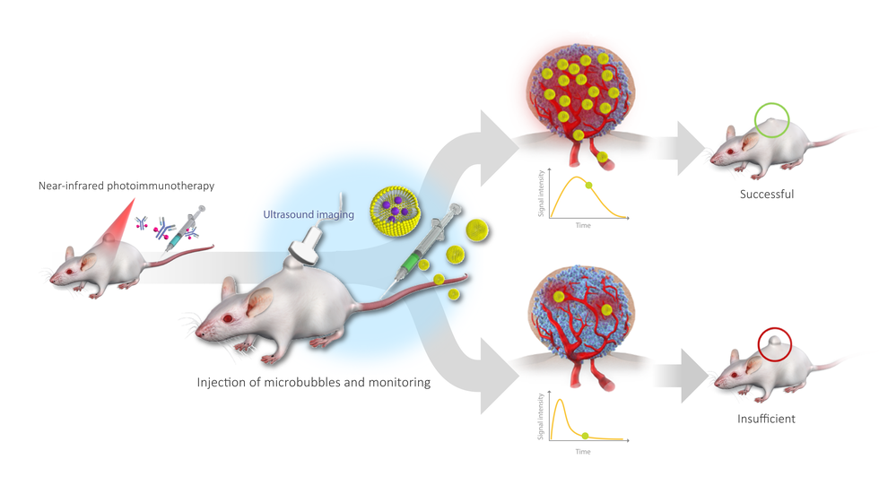
Researchers at Nagoya University in Japan and their collaborators have used a biomarker based on microbubbles to evaluate the success of near-infrared photoimmunotherapy (NIR-PIT) treatment. Using ultrasound to track the microbubbles, they were able to identify areas where cancer therapy had not been fully applied. Their findings suggest ways to improve NIR-PIT and make it a viable alternative treatment for various types of cancer.
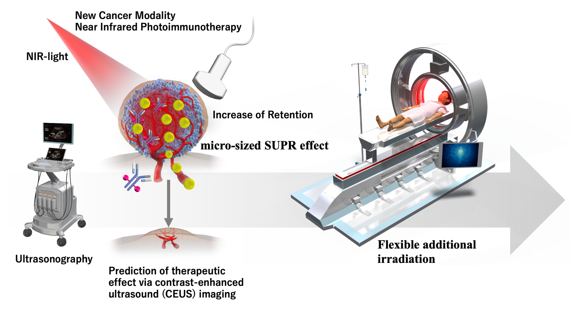
NIR-PIT is an innovative cancer treatment that combines the use of antibodies and near-infrared light to selectively destroy cancer cells while protecting healthy tissues. The antibody targets and binds to cancer cell proteins, creating a light-absorbing substance called IR700. When exposed to near-infrared light, IR700 activates and releases energy that destroys cancer cells. NIR-PIT is considered the fifth cancer treatment, along with surgery, radiation, chemotherapy, and cancer immunotherapy. Improving treatment could have important implications for cancer patients.
To effectively treat a tumor, physicians must determine the optimal level of light intensity to destroy abnormal cell growth while avoiding causing damage to healthy cells. However, it is difficult to ensure uniform irradiation of target cells during surgery because the host tissue reflects and scatters light. Since insufficient light irradiation to the whole tumor risks undertreatment, physicians need an indicator to judge its effectiveness.
To determine the best way to do this, Dr. Kazuhide Sato of Nagoya University Graduate School of Medicine and his collaborators looked at differences in tumor vessels compared to host cells. Past studies have reported that tumor vessels have irregular shapes, gaps between cells, and poor drainage. During NIR-PIT treatment, this poor drainage helps therapeutic nanoparticles stay in the tumor. They then enter cancerous tissue, resulting in a therapeutic effect known as the enhanced permeability and retention (EPR) effect.
In NIR-PIT treatment, rapid tumor cell death caused by the treatment increases permeability in the tumor vessels. This leads to a ‘super EPR effect’ (SUPR), an EPR effect that is 24-times higher than other therapies. If the SUPR effect occurs all over the tumor, it is likely that the treatment has been successful, whereas if it is isolated to certain regions, it is less likely that the treatment has been successful.
To assess this, they tested whether, using the increased permeability, larger fluorescent nanoparticles, such as Sonazoid microbubbles, could be retained. Microbubbles offer an easy way to measure the SUPR effect as they can be easily detected by reflecting harmonic signals off them.
“We investigated with larger-sized fluorescent particles of 2 mm and 5 mm sizes”, said Sato. “We found that retention increased with both sizes.” Using ultrasound imaging to track microbubbles, they created a novel biomarker that could measure the SUPR effect before and after treatment and evaluate the effectiveness of NIR-PIT. “In short, the higher the retention, the higher the anti-tumor effect of NIR-PIT,” he explained.
Sato hopes that their discovery will improve the treatment of cancer patients. “Using this new concept, we could confirm and predict the effects of the treatment after NIR light irradiation,” he said. “This is especially important for patients who receive insufficient treatment. Additional irradiation could be performed flexibly. Since ultrasound imaging equipment has already been introduced in most hospitals and the microbubble contrast agent used in this study has already been approved, this technology is easy to translate to the clinic.”
This technology is easy to translate to the clinic. Researchers hope that their discovery
will improve the treatment of cancer patients. (Credit: Kazuhide Sato)
This research involved collaboration with several institutions at Nagoya University, including the Graduate School of Medicine, the Institute of Advanced Research, the Graduate School of Engineering, and the Institutes of Innovation for Future Society, as well as the Institute for Quantum Life Science and JST.
The study, “Contrast-enhanced ultrasound imaging for monitoring the efficacy of near-infrared photoimmunotherapy,” has been published in the journal Ebiomedicine at DOI: 10.1016/j.ebiom.2023.104737.
Authors:
Kohei Matsuoka, Mizuki Yamada, Noriaki Fukatsu, Kyoichi Goto, Misae Shimizu, Ayako Kato, Yoshimi Kato, Hiroshi Yukawa, Yoshinobu Baba, Mitsuo Sato, and Kazuhide Sato
Media Contact:
Matthew Coslett
International Communications Office, Nagoya University
Email: icomm_research@t.mail.nagoya-u.ac.jp
Top image: Injected microbubbles were tracked by ultrasound imaging and used to evaluate the effectiveness of near-infrared photoimmunotherapy. (Credit: Kazuhide Sato)

