
Complex systems
July 14, 2022
AI analyses neuron changes to detect whether drugs are effective for neurodegenerative disease patients
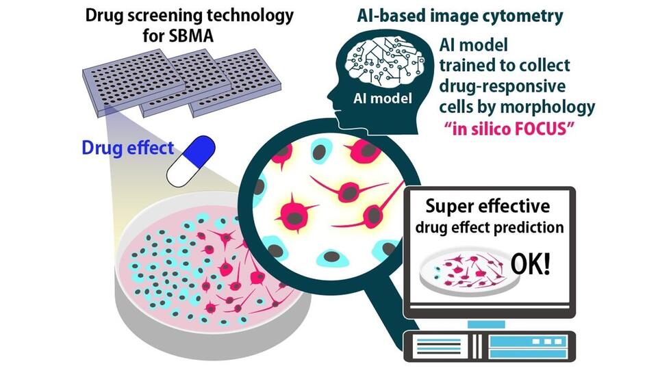
A research group from Nagoya University in Japan has developed an artificial intelligence for analyzing cell images that uses machine learning to predict the therapeutic effect of drugs. Called in silico FOCUS, this new technology may aid in the discovery of therapeutic agents for neurodegenerative disorders such as Kennedy disease.
Current treatments for neurodegenerative diseases often have harsh side effects, including sexual dysfunction and blocking muscle tissue formation. However, researchers searching for new, less harmful treatments have been hindered by the lack of effective screening technologies to discern whether a drug is effective. One promising concept is the ‘anomaly discrimination concept’, meaning neurons that respond to treatment have slight differences in shape compared to those that do not. However, these subtle differences are difficult to discern with the naked eye. Current computer technologies are also too slow to perform the analysis.
A group of Nagoya University professors, led by Associate Professor Ryuji Kato and Assistant Professor Kei Kanie of the Graduate School of Pharmaceutical Sciences, and Professor Masahisa Katsuno and Assistant Professor Madoka Iida of the Graduate School of Medicine, has developed a new artificial intelligence technology called in silico FOCUS. It analyzes the cell shape of model neurons and uses that information to assess whether they respond to therapeutic drugs. They published their results in the journal Scientific Reports.
The researchers tested the AI on a model of cells being treated for Kennedy disease, a neurodegenerative disorder that leads to motor neuron death. in silico FOCUS constructed a robust image-based classification model that had 100% accuracy in identifying the state of recovery of the model cells.
“This technology enables a highly sensitive and stable evaluation of the effects of therapeutic agents through the analysis of changes in the shape of diseased model cells to those of healthy cells, which we could not normally distinguish,” Professor Kato explains. “This is an ultra-efficient screening technology that can predict drug efficacy by simply capturing images, thus reducing the time required for drug efficacy analysis and evaluation from several hours with several hundred thousand cells to only a few minutes. It allows for a highly accurate prediction of therapeutic effects, without complicated and invasive experiments.”
Kato concludes: “These results suggest the possibility of accelerating the development of new drugs and we expect them to be widely applied to the discovery of therapeutic drugs for diseases that have been difficult to explore.”
Neuron analysis tool offers hope for research into neurodegenerative disorders
(Credit: Ryuji Kato)
The study, “Label-free morphological sub-population cytometry for sensitive phenotypic screening of heterogenous neural disease model cells,” was published in the journal Scientific Reports on June 16, 2022 at DOI: 10.1038/s41598-022-12250-0.
Authors:
Yuta Imai, Madoka Iida, Kei Kanie, Masahisa Katsuno, and Ryuji Kato
This research was supported by the FY2019 Nagoya University NU Cross-Departmental Innovation Creation Project.
Media Contact:
Matthew Coslett
International Communications Office, Nagoya University
Email: kouho-en@adm.nagoya-u.ac.jp

