
Interdisciplinary science and engineering
May 10, 2023
Glass filters developed to separate tumor cells from blood may increase efficiency of blood tests for cancer
A group from Nagoya University has identified the optimal dimensions of glass filters for circulating tumor cells (CTCs) and cancer-associated fibroblasts (CAFs), demonstrating that they could detect cells from lung cancer patients. Using a unique method, the group visualized the cells on glass filters and captured them. Their findings will enable more efficient culture of tumor cells to identify patients with cancer. The study was published in the journal Scientific Reports.
Early diagnosis is important to treat cancer. Relatively short periods of time in treatment can impact patient longevity and health. A promising approach is to detect CTC and CAFs shed by tumors in blood samples. Analyzing these cells provides direct information about cancer and can predict disease progression even in the early stages of the disease. However, the number of CTCs in the blood of cancer patients is extremely low. Therefore, improvements in the efficiency of capture and culture are necessary.
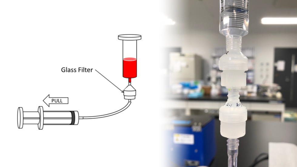
Glass is often used to culture cells in laboratories, but its use in CTC culturing remains underutilized. Therefore, a Nagoya University research team led by Professor Tanaka (he/him) of the Center for Low-Temperature Plasma Sciences, in collaboration with the Nagoya University Graduate School of Medicine and Institute of Innovation for Future Society, developed glass filters with a range of shapes and dimensions to determine which best captured CTCs and CAFs of lung cancer patients. They also developed a system that they called FROG-CHIP for visualizing the CTCs and CAFs captured on the glass filters.
The group found that the best patterns to capture CTCs while allowing white blood cells to pass through were a circular pattern with a pitch of 20 micrometers and a diameter of 8 micrometers. “These dimensions had high capture efficiency without retaining many white blood cells,” explained Tanaka.
“These results suggest that our glass filters are useful as a tool for the early diagnosis of tumors,” he continued. “Our findings would be useful at hospitals, clinics, and diagnostic testing companies to speed up cancer diagnosis.”
The study, "High-performance glass filters for capturing and culturing circulating tumor cells and cancer-associated fibroblasts," was published in the journal Scientific Reports at DOI: 10.1038/s41598-023-31265-9
Authors:
Hiromasa Tanaka, Daijiro Iwata, Yuki Shibata, Tetsunari Hase, Daisuke Onoshima, Naoyuki Yogo, Hirofumi Shibata, Mitsuo Sato, Kenji Ishikawa, Ikuo Nagasawa, Yoshinori Hasegawa, Makoto Ishii, Yoshinobu Baba, and Masaru Hori
Media Contact:
Matthew Coslett
International Communications Office, Nagoya University
kouho-en@adm.nagoya-u.ac.jp

