With the help of medaka fish, CRISPR and new imaging techniques, researchers have set a new standard for studying cell division at the very earliest stages of life.
メダカ、CRISPR、最新のイメージング技術を用い、生命の初期段階における細胞分裂の研究で新たな基準を打ち立てました。
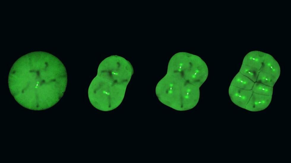
The beginning of life is shrouded in mystery. While the intricate dynamics of mitosis is well-studied in the so-called somatic cells ? the cells that have a specialized function, like skin and muscle cells ? they remain elusive in the first cells of our bodies, the embryonic cells. Embryonic mitosis is notoriously difficult to study in vertebrates, as live functional analyses and -imaging of experimental embryos are technically limited, which makes it hard to track cells during embryogenesis.
生命の誕生は謎に包まれています。有糸分裂の複雑な動態は、いわゆる体細胞(皮膚細胞や筋肉細胞のような特殊な機能を持つ細胞)ではよく研究されていますが、私たちの体の最初の細胞である胚細胞に関しては、いまだ解明されていません。脊椎動物における胚性有糸分裂の研究は困難であることが知られています。というのも、ライブイメージングでの機能解析が技術的に限られており、胚形成中の細胞を追跡することが難しいからです。
However, researchers from the Cell Division Dynamics Unit at the Okinawa Institute of Science and Technology (OIST) have recently published a paper in Nature Communications, together with Professors Toshiya Nishimura from Hokkaido University (previously at Nagoya University), Minoru Tanaka from Nagoya University, Satoshi Ansai from Tohoku University (currently at Kyoto University), and Masato T. Kanemaki from the National Institute of Genetics. The study takes the first major steps towards answering questions about embryonic mitosis, thanks to a combination of novel imaging techniques, CRISPR/Cas9 genome editing technology, a modern protein-knockdown system, and medaka, or Japanese rice fish (Oryzias latipes ). The timelapses that they have produced help answer fundamental questions about the intricate process of equally dividing chromosomes during embryonic mitosis, and simultaneously chart the next frontier of scientific exploration. As Professor Tomomi Kiyomitsu, senior author of the study, describes the timelapses: “they are beautiful, both on their own and because they lay a new foundation for elucidating embryonic mitosis.”
このほど、沖縄科学技術大学院大学(OIST)の細胞分裂動態ユニットの研究チームは、名古屋大学(現北海道大学)の西村俊哉助教、名古屋大学の田中実教授、東北大学(現京都大学)の安齋賢特定准教授、国立遺伝学研究所の鐘巻将人教授らと共に、学術誌『ネイチャーコミュニケーションズ』に論文を発表しました。本研究は、新しいイメージング技術、CRISPR/Cas9ゲノム編集技術、最新のタンパク質ノックダウンシステム、そしてモデル生物としてミナミメダカ(Oryzias latipes )を用いることによって、胚性有糸分裂の解明のための、最初の大きな一歩となりました。研究チームが作成したタイムラプスは、胚で染色体が均等に分かれる複雑なプロセスについての根本的な理解に役立つとともに科学的探求の次のフロンティアにつながります。本研究の責任著者である清光智美准教授は、タイムラプスについて次のように語っています。「タイムラプスの画像自体も美しいのですが、胚の有糸分裂の解明につながる新たな基礎を築いた点も評価できます。」
◆詳細(プレスリリース本文)はこちら